Prof. Jun Liu Published article on ACS Appl. Mater. Interfaces with the title of "Development of Cellulose Nanofibril/Casein-Based 3D Composite Hemostasis Scaffold for Potential Wound-Healing Application"
ABSTRACT: Excessive bleeding in traumatic hemorrhage is the primary concern for natural wound healing and the main reason for trauma deaths. The three-dimensional (3D) bioprinting of bioinks offers the desired structural complexity vital for hemostasis activity and targeted cell proliferation in rapid and controlled wound
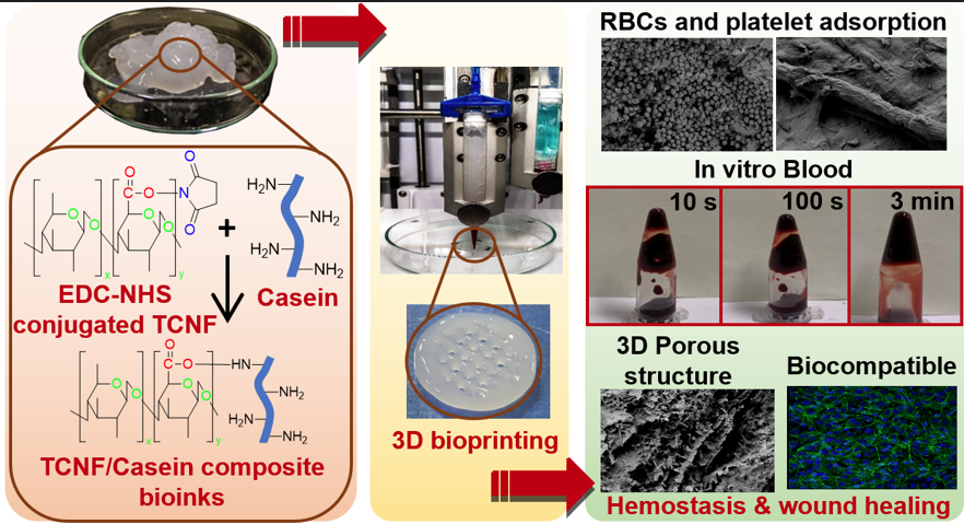
healing. However, it is challenging to develop suitable bioinks to fabricate specific 3D scaffolds desirable in wound healing. In this work, a 3D composite scaffold is designed using bioprinting technology and synergistic hemostasis mechanisms of cellulose nanofibrils (TCNFs), chitosan, and casein to control blood loss in
traumatic hemorrhage. Bioinks that consist of casein bioconjugated TCNF (with a casein content of 104.5 ± 34.1 mg/g) using the carbodiimide cross-linker chemistry were subjected to bioprinting for customizable 3D scaffold fabrication. Further, the 3D composite scaffolds were in situ cross-linked using a green ionic
complexation approach. The covalent conjugation among TCNF, casein, and chitosan was confirmed by Fourier transform infrared (FTIR) spectroscopy, nuclear magnetic resonance (NMR), X-ray photoelectron spectroscopy (XPS), sodium dodecyl sulfatepolyacrylamide gel electrophoresis (SDS-PAGE), and X-ray diffraction (XRD) studies. The in vitro hemostasis activity of the 3D composite scaffold was analyzed by a human thrombin−antithrombin (TAT) assay and adsorption of red blood cells (RBCs) and platelets. The 3D composite scaffold had a better swelling behavior and a faster whole blood clotting rate at each time point than the 3D TCNF scaffold and commercial cellulose-based dressings. The TAT assay demonstrated that the 3D composite scaffold could form a higher content of thrombin (663.29 pg/mL) and stable blood clot compared to a cellulosic pad (580.35 pg/mL), 3D TCNF (457.78 pg/mL), and cellulosic gauze (328.92 pg/mL), which are essential for faster blood coagulation. In addition, the 3D composite scaffold had a lower blood clotting index (23.34%) than the 3D TCNF scaffold (41.93%), suggesting higher efficiencies for RBC entrapping to induce blood clotting. The in vivo cytocompatibility was evaluated by a 3D cell culture study, and results showed that the 3D composite scaffold could promote growth and proliferation of NIH 3T3 fibroblast cells, which is vital for wound healing. Cellulase-based in vitro deconstruction of the 3D composite scaffold showed significant weight loss (80 ± 5%) compared to the lysozyme hydrolysis (22 ± 5%) after 28 days of incubation, suggesting the biodegradation potential of the composite scaffold. In conclusion, this study proposes efficient prospects to develop a 3D composite scaffold from bioprinting of TCNF-based bioinks that can accelerate blood clotting and wound healing, suggesting its potential application in reducing blood loss during traumatic hemorrhage.
KEYWORDS: hemostasis, bioprinting, bioinks, nanocellulose, wound healing, scaffold
https://doi.org/10.1021/acsami.1c21039